4236099
Transport in mammals
Description
Slide Set by Aarushi Pandit, updated more than 1 year ago
More
Less
|
|
Created by Aarushi Pandit
over 8 years ago
|
|
Resource summary
Slide 1
Transport In Mammals
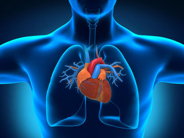
Caption: : Location of the human heart in the thoraic cavity. (Notice that the left lung is smaller to adjust the heart)
Slide 2
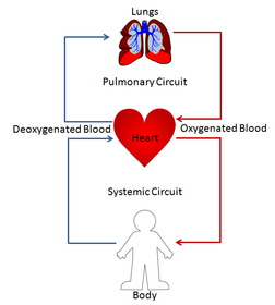
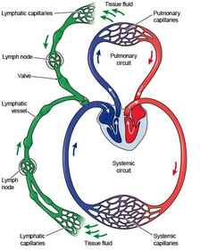
Pulmonary and Systemic Circulation
Pulmonary circulation:It is the portion of the cardiovascular system which carries deoxygenated blood away from the heart, to the lungs, and returns oxygenated (oxygen-rich) blood back to the heart.
Systemic circulation:It is the part of the cardiovascular system which carries oxygenated blood away from the heart to the body, and returns deoxygenated blood back to the heart.
Slide 3
Caption: : A very simplified depiction of pulmonary and systemic circulation
Slide 4
A closed double circulatory system
The combination of pulmonary circulation and systemic circulation causes the blood to travel twice to the heart in one complete circuit, all the while flowing in defined, closed vessels. Thus, the mammalian circulatory system can be termed a closed double circulatory system.
Slide 5
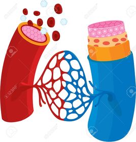
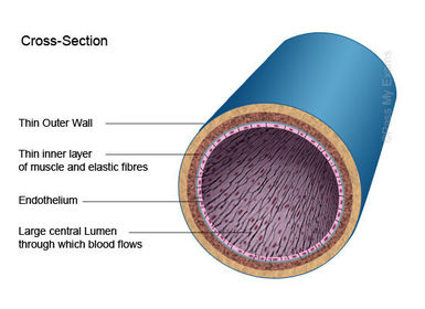
Blood Vessels
Caption: : Arteries (thick, red) Capilaries (thin, blue and red) Veins (thick, blue)
Slide 6
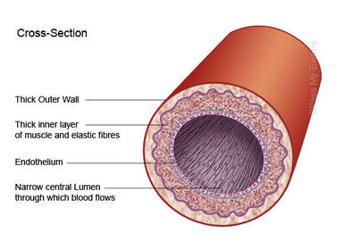
Arteries
Slide 7
Capillaries
Slide 8
Veins
Slide 9
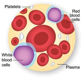
Blood Plasma and Tissue Fluid
Blood is composed of cells floating in a pale yellow liquid called plasma. As blood flows through capillaries within tissues, some of the plasma leaks out through the gaps between the cells in the walls of the capillary, and seeeps into the spaces between the cells of the tissues. This leaked plasma is known as tissue fluid.Tissue fluid is almost identical in compostion to blood plasma. However, it contains far few protein molecules than blood plasma, as these are too large to escape easily through the tiny holes in the capillary endothelium. Red blood cells (RBCs) are too large to pass through, so tissue fluid does not contain these, but some white blood cells (WBCs) can squuze through and move around freely in the tissue fluid.
Slide 10
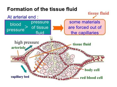
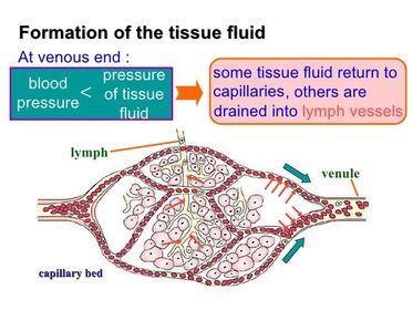
Slide 11
Oedema
If the blood pressure is too high, too much fluid is forced out of the capillaries, and may accumulate in the tissues.This build-up of fluid is called Oedema.
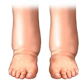
Slide 12
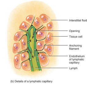
Lymphatics
About 90% of the fluid that leaks from the capillaries, seeps back into them. The remaining 10% is collected up and returned to the blood system by means of a series of tubes known as lymph vessels or lymphatics.Lymphatics are tiny, blind-ending vessels, which are found in almost all tissues of the body. They contain tiny valves, which allow the tissue fluid to flow in but stop it from leaking out. These valves are wide enough to allow large protein molecules to pass through. This is very important because such molecules are too big to get into blood capillaries, and so cannot be taken away by the blood.If your lymphatics did not take away the protein in the tissue fluid between your cells, you could die within 24 hours!!
Slide 13
Caption: : Notice the valves here
Slide 14
The fluid inside the lymphatics is called lymph. Lymph is virtually identical to tissue fluid; it has a different name more because it is in a different place than because it is different in compostion. In some tissues, the tissue fluid, and therefore the lymph, is rather different from that in other tissues.
Lymph
Caption: : lymph
Slide 15
Lymphatics join up to form larger lymph vessels, that gradually transport the lymph back to the large veins that run just beneath the collar bone, the subclavian veins.As in veins, the movement of fluid along the lymphatics is largely caused by the contraction of muscles around the vessels, and kept going in the right direction by valves.Lymph vessels have smooth muscle in their walls, which can contract to push the lymph along. Lymph flow is very slow.
Slide 16
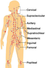
Caption: : Lymph nodes (in red) throughout the human body
At intervals along lymph vessels, there are lymph nodes.These are involved in protection against disease. Bacteria and other unwanted particles are removed from lymph by some types of WBCs as the lymph passes through a node, while the other white blood cells within the nodes secrete antibodies.
Lymph Nodes
Slide 17
Blood
Slide 18
Haemoglobin
Oxygen is transported around the body inside red blood cells in combination with the protein haemoglobin.Haemoglobin + Oxygen -> OxyhaemoglobinOxyhaemoglobin -> Haemoglobin + OxygenEach haemoglobin molecule is made up of four polypeptides, each containing one haem group. each haem group can combine with one oxygen molecule, O2. Overall, then each haemoglobin molecule can combine with four oxygen molecules (eight oxygen atoms).
Slide 19
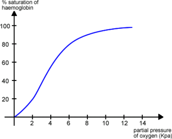
The haemoglobin dissociation curve
To investigate how haemoglobin behaves, samples are extracted from blood and exposed to different concentrations, or partial pressures of oxygen.The amount of oxygen that combines with each sample of haemoglobin is then measured.A sample of haemoglobin which has combined with the maximum possible amount of oxygen is said to be saturated.The percentage saturation of each sample can be plotted against the partial pressure of oxygen to obtain the curve known as the dissociation curve.
Slide 20
Caption: : The haemoglobin dissociation curve
Consider thehaemoglobin within a RBC in a capillary in the lungs. Here the partial pressure of Oxygen is high, therefore this haemoglobin will be 95-97% saturated with oxygen.In an actively respiring muscle, on the other hand, where the partial pressure of oxygen is low, the haemoglobin will be about 20-25% saturated with oxygen.From this we can infer that haemoglobin coming from the lungs carries a lot of oxygen; as it reaches a muscle, it releases around three-quarters of it.
Slide 21
The S-shaped curve
The shape of the haemoglobin dissociation curve can be explained by the behaviour of a haemoglobin molecule as it combines with or loses oxygen molecules.When an oxygen molecule combines with one haem group, the whole haemoglobin molecule is slightly distorted. The distortion makes it easier for a second oxygen molecule to combine with a second haem group. This in turn makes it easier for a third oxygen molecule to combine with a third haem group. It is then still easier for a fourth and final oxygen molecule to combine.Up to an oxygen partial pressure of around 2kPa, on average only one oxygen molecule is combined with each haemoglobin molecule. Once this oxygen molecule is combined, however, it becomes succesively easier for the second and third oxygen molecules to combine, so the curve rises very steeply. Over this part of the curve, a small change in the partial pressure of oxygen causes a very large change in the amount of oxygen which is carried by the haemoglobin.
Slide 22
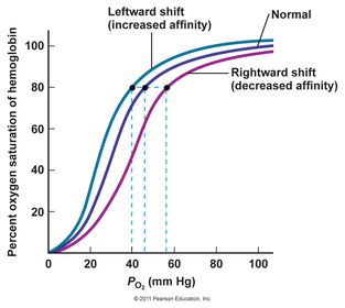
Bohr Shift
Caption: : Left shift: low CO2 conc. Right shift: high CO2 conc.
The amount of oxygen the haemoglobin carries is affected not only by the partial pressure of oxygen, but also by the partial pressure of carbon dioxide (CO2).The presence of a high partial pressure of carbon dioxide causes haemoglobin to release oxygen. This is called the Bohr effect, after Christian Bohr who discovered it in 1904.
Slide 23
Bohr shift (continued..)
High concentrations of carbon dioxide are found in actively respiring tissues, which need oxygen; these high carbon dioxide concentrations cause haemoglobin to release its oxygen even more readily than it would otherwise do. At each partial pressure of oxygen, the haemoglobin is less saturated than it would be a low partial pressure of carbon dioxide.
Slide 24
Problems with oxygen transport
Slide 25
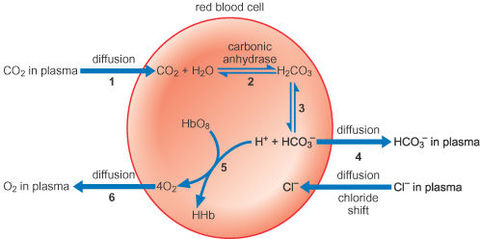
Carbon dioxide transport
Caption: : 80% of CO2 is transported as dissolved hydrogencarbonate ions, 10% as carbaminohaemoglobin and 5% dissolves as it is in blood plasma.
Slide 26
The heart
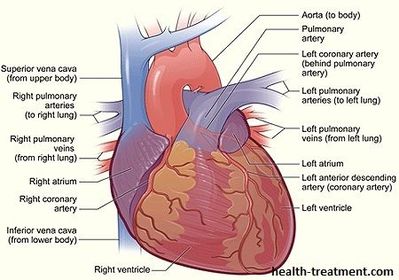
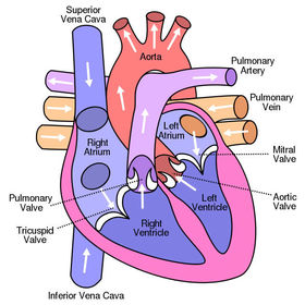
Slide 27
The Cardiac Cycle
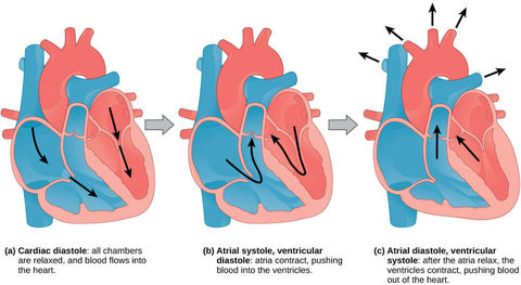
Slide 28
How the heart valves function
Caption: : blue arrow indicates the direction of the pressure.
Slide 29
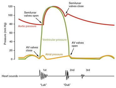
Pressure changes during the cardiac cycle
Slide 30
Control of the heart beat
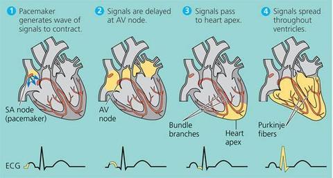
Each cardiac cycle begins in the right atrium. There is a small patch of muscle tissue in the right atrium wall known as the sinoatrial node (SAN), which automatically contracts and relaxes all the time. It does not need a nerve impulse to start it off, so it is said to be myogenic- that it is 'started by the muscle'.The pacemaker's rate can be adjusted by nerves transmitting impulses to the pacemaker from the brain
Slide 31
As the muscle in the SAN contracts, it produces an electrical excitation wave which sweeps through all of the muscle in the atria of the heart. This excitation wave makes the atrial walls contract.The excitation wave sweeps onwards and reaches another patch of cells, called the atrioventricular node (AVN). This node is the only way in which electrical impulses can get down to the ventricles. The AVN delays the impulse for a fraction of a second before it travels down into the ventricles. This delay means that the ventricles receive signal to contract after the atria receive the signal.
Control of the heart beat (continued..)
Slide 32
Control of the heart beat (continued..)
The excitation wave moves swiftly down through the septum of the heart, along fibres known as Purkyne tissue. Once the excitation wave arrives at the base of the ventricles it sweeps upwards, through the ventricle walls. The ventricles contract.The ventricles then relax. The muscle in the SAN contracts again, and the whole sequence runs through once more.
Slide 33
Control of the heart beat (continued..)
Caption: : Purkyne tissue is also called Purkinje fibres.
Slide 34
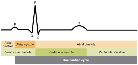
Electrocardiograms (ECGs)
Slide 35
Electrocardiograms (ECGs)
In the diagram shown before:The part labelled P represents the wave of excitation sweeping over the atrial walls.The parts labelled Q, R and S represent the wave of excitation in the ventricular walls.The T section indicates recovery of the ventricular walls.
Want to create your own Slides for free with GoConqr? Learn more.